I could not find the exact year in which the skin sample you are about to see was tanned. It's not that I can't come up with the year, I just couldn't find anything handy to get the date off of.
Regardless, it was from either the '97, '98, or '99 season, which would make it well over one year before the time of this writing.
The skin sample was from a bow kill buck that was not found until the next day. A big un', even had B&C antlers on top of his head. A buddy of mine got the taxidermy job, IF he could save the cape.
I did not tan this cape, but I gave instructions over the phone as what to get, and what to compound, and how to use.
This cape was not salted before going into the pickle. As far as the pickle, neither salt as sodium chloride or potassium chloride were used. However, the pickle was a high SULPHATE pickle.
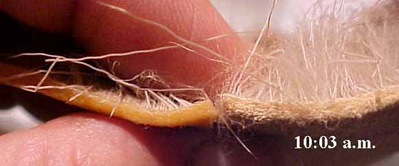
The first photo is of the sample right after I pulled it out of the cabinet where it had been stored over the years with temperatures that ran from the mid sixties to low nineties F.
One of the down sides of the treatment I had this cape subjected to is that the acid will oxidize or burn off the brown pigments that are on the outside of the hair shaft. Part of the pigments that make up the color of a deer's hair are also on the "inside" of the hair. In this case browns were oxidized, leaving a red look. Some can wind up with a gray look.
In either case, the average person would most likely not know the difference. Just a fact of life.
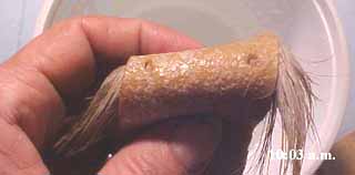
Here's the flesh side before I whack a piece off.
Notice the time is marked on all the photos.
I have put the skin piece I whacked off in a bowl of tap water with a pH of a ballpark of 8. NO SALT was added.
The skin is soaked up, notice the amount of elapsed time. How could it do that that fast?
I am holding the cut piece next to where I had cut if off at. How's come there's no acid swell?
Just another photo of the flesh side. It shows a couple of holes where I had taken samples from in the past for microscope work. To date, basically nothing has changed.
What I'm showing in this photo is a pH strip laid on the flesh side that gives a reading of the effluent coming out of the skin. Remember, the tap water was a pH of 8, with no salt.
Why didn't this piece disintegrate?
Why doesn't it have acid swell?
It's obviously acid, and indications would be that it would be of a sulphuric nature.
I cut off a cross section of skin. I could not tease it apart with straight pins. I had to use dental tartar scrapers to separate it enough to get these photos of the collagen fiber structures.
The following photos were all from the same field of view. They are both top light, and back light. Pay attention to the time frames and the behavior of the collagen fibres.
How and why do the fibres appear to be moving and changing in shape?
The last photo is a masterpiece. If it's fact, it ain't braggin'.