MICROSCOPE PHOTO HAIR FOLLICLE
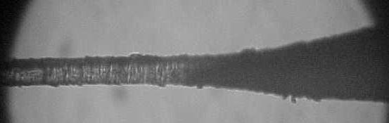
This view shows the long, skinny section of the hair shaft where it joins to the hollow portion. You see that it tapers into a flare before becoming cylindrical in structure.
Notice the shaft appears to be an assemblage of plate-like structures, not unlike a stack of coins. At this time I really do not know if that structure is bonded any better than what it appears, or if there is a more solid "core like" structure underneath the visible. My suspicion is.......what you see is what you got.
MICROSCOPE PHOTO HOLLOW PORTION MAGNIFIED
Beyond the Senses
Illustrated with Photos Taken Through the Lens
of a Microscope and Camera
An indepth study by Glen Conley
HAIR STRUCTURE AND HAIR SLIP
The more I learn, the more I realize how little I know. Do you ever have that problem? Or is it really even a problem? Maybe it is just something programmed into us to keep us humble. For every answer there is a dozen questions. The more we see the more we learn, the more we learn the more we see. Never ending cycle. But it can be fun and entertaining, but it can also be exasperating and frustrating.
Just like any normal twelve year old would do, I begged my parents for a microscope for Christmas that year. I got the 'scope plus a bonus, two text books. While my abnormal brother and sister stayed plugged into the T.V., I stayed plugged into the microscope.
I learned early in life that there was a "world" beyond our senses. Come with me on a short trip into this world. I would be willing to bet that it will change the way you see the obvious.
The specimen samples were acquired from five different whitetail deer. If you have been further confused in the past with Folk-artsy pen and ink washes for illustration, feel relieved, these are all actual photos.
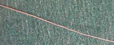
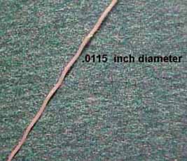
STRAIGHT DEER HAIR
The caption for this photo pretty well says it all.
ZIG-ZAGGY DEER HAIR
Contrast this hair structure with the straight hair. Amazing difference isn't there? It is obvious that the "coded sequence" for the structure of this hair is "written" differently than that of the straight hair.
I had been working in such a narrow range of focus that it took me an embarassingly long amount of time to make the correlation of a predisposition to hair slip with the zig-zaggy hair. I couldn't see the forest for the trees. That comment was not a dyslexia attack.
Once my dim light bulb finally came on, I also realized that there was also a structural difference to the skin associated with different hair types. Prior to seeing the light, I had worked at the microscopic level with the skin and hair as two separate entities. Looking back, I can say, "That was dumb".
You want to know what's really bad about this lack of observation and correlation? I am involved with a rare breed of horses. Curly horses. If you ever even heard of the breed, it is probably due to comparatively recent publications.
A number of years ago I was actively involved in Curly horse research, but I was working more on back-tracking historically. University geneticists and biologists were also involved.
The first areas the biologists "attacked" were the hair, then the skin, then the FAT deposits to differentiate the Curlies from other horses. (digestive tract and skeletal structure were also worked on F.Y.I.) In essence, some of the obvious answers to my questions had been right in front of my eyes, every day of the week for years! I just didn't think to use the Curlies' attributes as an analogy for whitetail skin and hair structure even though I used horse and whitetail skeletal and musculature analogies on a regular basis in taxidermy work.
As you go through this series, don't make the same mistake I did. Keep your eyes wide open and your mind and memory working. Remember your own past life experiences and observations. See the forest, then the trees and then the leaves of the trees and then come back to look at the forest again.
TINE INJURY SAMPLE
This sample was taken from the edge of a tine wound on the side of the neck. It was right at the edge of the scab. I had soaked the scab and area adjacent to with water. That allowed this sample to come right off.
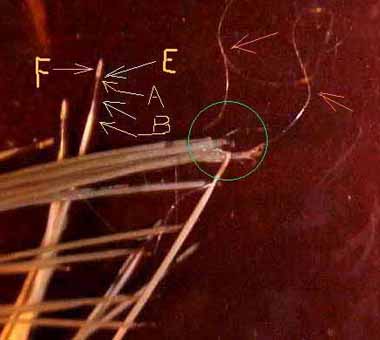
This will give you an idea as to what a "normal" hair will look like if you put your first attention on the hair that has the F designation. The F is the follicle.
The A and the next arrow down, points out the shaft.
B points out the hollow part of the hair.
Compare the E area to the enlarged photo of a follicle.
The two arrows that point to the silvery threads, those silvery threads are under coat.
You might want to come back and take another look at the area that is circled, once you have gone through this article. By that time, you should have an understanding as to what has taken place.
Notice that these hairs are straight.
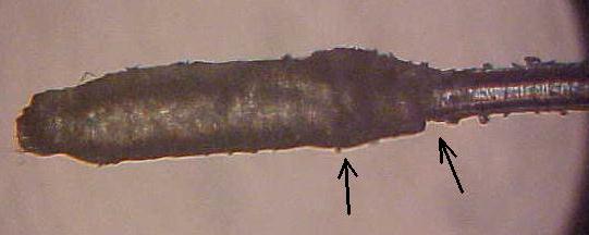
This photo will give you a pretty good idea of what the hair follicle looks like. This photo will also give you a pretty good idea how to think like a detective. The two arrows identify the evidence.
A hair sample was sent to me by a beginning taxidermist that was having trouble with the hair slipping from his capes after they were mounted. The arrow next to the shaft identifies a tear. The second arrow identifies bunching up around the enclosed section of shaft.
This hair had been physically pulled from the cape. The tearing and bunching were caused by the follicle being pulled through the epidermis. Just like pulling a root vegetable like a carrot or radish from the soil. Ya reckon that's where the term "hair roots" came from?
I was not familiar with the products that this taxidermist used to prepare his capes. Hairs like this will normally be seated pretty good, once the leather has dried. The taxidermist confirmed at a later date that the hair was "tight" once the mounts were thoroughly dry.
In a live animal the various structural strata of the skin can "slide", to a degree, back and forth and in and out across each other.
You can demonstrate this to yourself. Take your thumb and finger and pinch up a section of skin on the side of your neck. Now pull it out and roll it between your thumb and finger.
If some one walks into the room at this point and sees you staring into your monitor and pulling the skin on your neck, they are probably going to ask, "What in thee Hell are you doing?" They probably really don't want to know, they are just asking you that question in an effort to make you feel stupid.
Give them this answer, "I am demonstrating to myself the ability of the epidermis, dermis, and the collagen fibers contained with in the dermis, and the hypodermis to move together in a degree of unison or a degree of independent horizontal movement across each other."
That will probably make them feel stupid, and they will leave you alone to study in peace. Once out of sight, they are probably pulling on their own neck skin tryin' to figure this out so they don't have to feel so stupid.
If you and your observer were to do this same exercise with the neck skin of a live deer, you both would probably notice the feel of the skin between your thumbs and fingers to be remarkably similar to your own skin. Don't be surprised if the deer's skin is more elastic.
A desirable to work with skin for taxidermy will have a degree of elasticity, or "stretch" and not a static "memory". An acid pickle has dissolved a lot of the "working bonds" that the live skin had, and as a result, being able to pull hairs out of a wet, freshly mounted cape is not uncommon. I might also add that the state of decomposition that the cape was in prior to being worked with is always an unknown factor we have to work with or around.
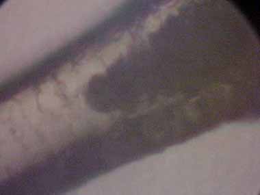
MICROSCOPE PHOTO SHAFT/HOLLOW PORTION UNION
MICROSCOPE PHOTO UNION AREA MAGNIFIED
The dark solid color "V" shape that you see here is pigment contained INSIDE the beginning of the hollow portion of the hair. This illustrates why a whitetail cape can not be bleached out to white.
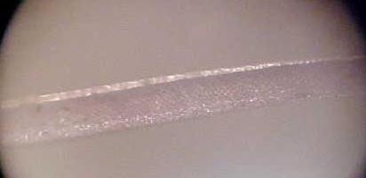
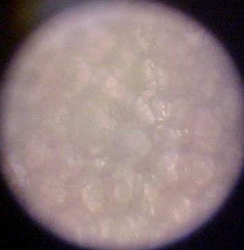
MICROSCOPE PHOTO HOLLOW PORTION
This photo is of the hollow portion of the hair. You will notice the light colored areas that parallel the outer edges of the hair shaft and the darker area in between. The light areas are the walls of the cylindrical structure. Remember the eleven and one half thousandths of an inch measurement you saw earlier? That was the diameter of the hair pictured. This gives you a real good idea as to how thin those walls are.
I took this picture with a magnification of 1750x. It's going to be a lot more than that on your computer monitor!
This shows the hollow bubble like structure of the hollow portion of hair. The darker, low areas contain pigment. I would have liked to have had a hair from a raw skin that had a higher degree of darker pigment for this photo, but a feller always has to work with what he's got.
By the way, many bacteria will show up well enough at 600x-650x that they are identifiable by shape.
Rod shaped bacillus can be differentiated from round shaped staphlyococcus, and round shaped streptococcus can be distinguished from the staph.
Some of the most important photos explaining hair slip in whitetail deer are found on the next page. These are photos of slip conditions like you were probably unaware of until now.
Continue to page two of Beyond the Senses and more microscope photos.